 |
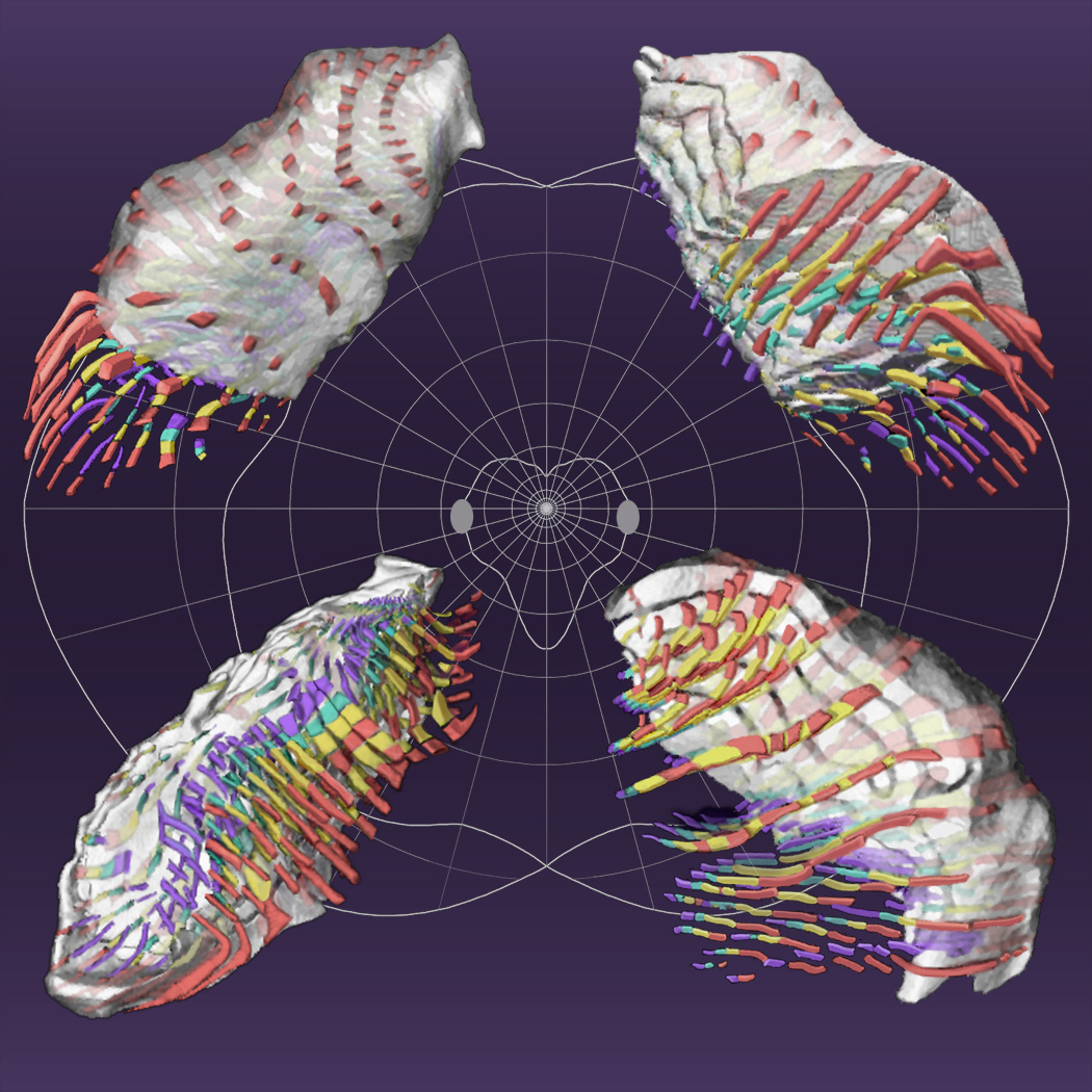
Views of the lateral geniculate nucleus of the rhesus monkey extracted
from a functional atlas.
Projection columns representing directions in visual space: red, contra-eye
parvo; yellow, ipsi-eye parvo; green, ipsi-eye magno; blue, contra-eye magno.
Columns are retinotopically located at intersections of the light grey
background polar grid (more clearly visible on the
high-resolution image): inclinations every 15°;
eccentricities spanning roughly equal distances in the nucleus (0°,
1.0°, 1.9°, 3.4°, 6.3°, 11.3°, 19.2°, 30.7°, 46.6°,
and 67.6°). The heart-shaped central figure intersecting the oval blind spots
separates regions of visual space represented by six (central) and four (peripheral)
geniculate layers. Cut faces of semitransparent views pass though the blind spot:
upper panels show the 17° isoeccentricity surface; lower panels show the minus
7° isoinclination surface. The posterior pole (foveal representation) is
in the uppermost part of each panel; the lower-left panel shows the ventral
surface. Projection columns are retouched to improve clarity. Made with the
assistance of Janet Sinn-Hanlon of the
Visualization, Media and Imaging Laboratory of the Beckman Institute for Advanced
Science and Technology, University of Illinois at Urbana-Champaign.
|